| Parasite | Microsporidium cypselurus |
|---|---|
| Taxonomy | Microspora, Microsporea |
| Host | Bennett’s flyingfish (Cypselurus pinnatibarbatus) |
| Infection site | Trunk muscle |
| Clincal sign |
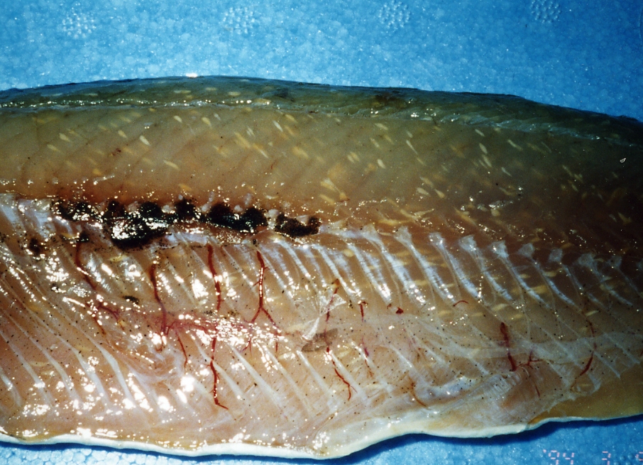
Small aggregates of cysts (max. 3-4 mm) are observed in the muscle (Fig. 1). Thecysts are spindle-shaped and white in colour. |
| Parasitology | Many spores are produced inside the cysts (Fig. 2). The spores (length 3.7-4.8 (average 4.1) mm; width 2.1-2.7 (2.2) mm) are ovoid to pyriform in shape. The presence of sporophorous vesicles has not been clearly demonstrated. |
| Pathology | The cysts are encapsulated by the host-produced thin fibrous connective tissues. The myoliquefaction does not occur (Yokoyama et al., 2002). |
| Health hazard | Since this parasite is not infectious to human, it is harmless in food hygiene. |
| Diagnosis | Check the spores by wet-mount of cysts. Sample should be smeared and stained by Uvitex 2B followed by a fluorescent microscopic observation. The stained spores emit blue fluorescence under UV radiation. |
| Other information | This microsporidium was observed in one individual of the Bennett’s flyingfish from Yakushima (Yokoyama et al., 2002). |
| References | Yokoyama, H., S. J. Lee and A. S. Bell (2002):
Occurrence of a new microsporidium in the skeletal muscle of the flying fish Cypselurus pinnatibarbatus japonicus (Exocoetidae) from Yakushima, Japan. Folia Parasitol., 49, 9-15. |
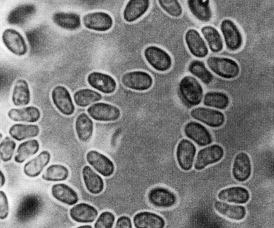
Fig. 1. Many microsporidian cysts in the trunk muscle of flying fish
Fig. 2. Fresh spores of M. cypselurus